檢測認證人脈交流通訊錄
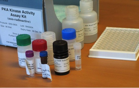
蛋白激酶A活性檢測ELISA試劑盒
- 這真不是您需要的產品?
- 品 牌:
- immuneChem
- PKA Kinase Assay Kits, Type I
Kits Components
1. PKA substrate plate: a 96 well plate with 12x8 removable strips. Each well was pre-immobilized with 50 μL of PKA-substrate (KRREILSRRPSYR).
2. Anti-pSubs (pCreb) antibodies (rabbit, affinity purified): 1 μg/mL, 5 mL.
3. Kinase Assay Dilution buffer: 10 mL.
4. Antibody dilution buffer: 10 mL.
5. Goat anti-rabbit IgG HRP conjugates: Catalog#ICP9803, 0.5 mg/mL in HRP stablizing buffer, 30 μL. Dilute to 0.5 μg/mL (1:5000 dilution) with antibody dilution buffer (D) as working solution.
6. Adenosine tri-phosphate (ATP): 2 mg. Reconstitute with 2 mL kinase assay dilution buffer (C) as working solution. Once it was dissolved in the buffer, store at -20 ℃ for better stability.
7. Peroxidase substrate: 10 mL, TMB (3,3'-5,5'-Tetramethylbenzidine).
8. Stop solution: 10 mL.
9. 10xPBSt: 10 mL.
Kits Description
1. Quality control: The kits were tested using PKA from Sigma (#P5511). Sensitivity is approximately 100 ng of PKA/assay.
2. Storage and Stability: Stable for 6 months at 4 ℃ for date of shipment. Avoid the light and heat.
3. Descriptions: PKA is a cyclic AMP dependent protein kianse. The PKA kinase assay kit (Type I) is a non-radioactive, homogenous, simple, rapid, antigen-captured immunosorbent assay. This assay is designed for the assay of PKA in solution. The principle of the assay is simple. Purified or partially purified PKA will phosphorylate the PKA substrate on the 96-well plate with the presence of ATP. The phosphorylated substrate (pSubstrate) is then analyzed with the anti-phosphosubstrate antibodies. Subsequently, the binding anti-pSub antibodies will be quantified with a secondary antibody-HRP conjugates, which generate the color signal from its substrate, TMB (OD at 450 nm). The final color intensity is proportional to the initial PKA phosphorylation activity.
Summary of the Assay
1. Kinase reaction:
KRREILSRRPSYR + ATP+Kinase → KRREILSRRPpSYR
2. The phosphorylated substrate on the plate will be detected with an anti-pSubstrate antibody:
KRREILSRRPSYR (no antibody binding)
KRREILSRRPpSYR (antibody binds to the plate)
3. Signal Generation: Second antibody binds to antibody-KRREILSRRPpSYR complex formed in the plate.
Protocol
1. Soak the well of the substrate plate (component A) with 50 μL of kinase assay dilution buffer (component C) for 10 mins, then aspirate.
2. Prepare ourified or partially purified PKA or PKA-inhibitor mixture in 30 μL of the kinase assay dilution buffer to the well of the substrate plate as prepared in step 1 (Note: For preparation of the positive control, refer to the Appendix)
3. Add samples to the appropriate wells.
4. Add 10 μL of the ATP (component F) solution to the well to initiate the kinase reaction at 30-35 ℃ for 90 mins. Hand-shake the strip well in every 20 mins. If a shaker with rotate angle is applied, the speed arranged at 60 rounds per min should be adequate.
5. Empty the well then add 40 μL of the anti-pSubstrate antibodies (component B ) at room temperature for 60 mins. shake the well in every 20 mins.
6. Empty the well and fill the well with 100 μL of PBSt wahsing buffer. Let the washing buffer stay for 1-2 mins then aspirate. Repeat this washing step for four times.
7. Add 40 μL of goat anti-rabbit IgG HRP (component E ) working soluiton to the well and incubate at room temperature for 30 mins. Shake the well in every 10 mins.
8. Empty the well and repeat the washing as step 6.
9. Add 60 μL of TMB to develop the color (for 30-60 mins).
10. Stop the reaction with 20 μL of the stop solution (2 N HCL).
Calculation of the relative Kinase Activity
OD(smaple)-OD(negative)/ng of kinase
Quality Control Test
Assay for activity of PKA (Sigma#P5511, lot#108H7846)
Activity, transfer 1 picomole of phosphate to kemptide/min/μg of protein.
Appendix
Preparation of Positive Control
Sample Preparation of Recombinant Kinase
1. Label up 5 microfuge tubes; 1-4, plus a negative control. Note: The negative control should only contain assay dilution buffer; no kinase.
2. Aliquot 38 μL of assay dilution into tube#1 and 30 μL into tubes 2-4.
3. Add 2 μL of recombinant kinase into tube#1 and mix thoroughly.
4. Transfer 10 μL from tube#1 to tube#2 and mix.
5. Transfer 10 μL from tube#2 to tube#3 and mix.
6. Transfer 10 μL from tube#3 to tube #4 and mix.
7. Transfer 30 μL from each of the mixtures (tubes 1-4 plus the control) into the appropriate wells of the substrate plate.
主站蜘蛛池模板:
亚洲欧美精品在线观看|
国产欧亚日韩视频|
97欧美精品一区二区三区|
欧美 日韩 国产 在线观看|
久久精品久久精品亚洲人|
狠狠色综合欧美激情|
国产精品亚洲二区在线观看|
99久久久精品免费观看国产|
日韩中文字幕在线不卡|
日韩精品―中文字幕|
欧美亚洲黄色片|
国产亚洲综合视频|
亚洲综合最新在线|
久久免费福利视频|
日韩欧美99|
亚洲爆乳无码专区|
91精品国产综合久久久久久久久|
国产在线精品播放|
久久另类ts人妖一区二区|
日韩av不卡播放|
日韩中文字幕精品|
色综合久久久久久中文网|
中文字幕日韩精品一区二区
|
精品国产综合久久|
日韩av第一页|
亚洲免费视频一区|
日韩中文在线视频|
日韩视频免费中文字幕|
亚洲欧洲精品一区|
亚洲午夜高清视频|
亚洲欧洲国产精品久久|
亚洲精品女av网站|
伊人久久大香线蕉成人综合网|
中文精品一区二区三区
|
日韩成人av电影在线|
一区二区不卡在线|
午夜久久资源|
欧洲精品久久久|
久久久久国产精品熟女影院|
久久国产精品网站|
国产精品亚洲二区在线观看|